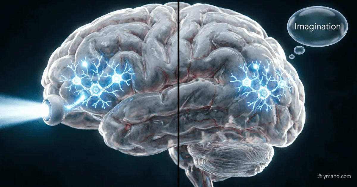
Unified Neural Mechanism for Imagination and Real Vision Discovered
Scientists at Cedars-Sinai have proven that imagining objects and actually seeing them engage the same neurons in the brain. About 40% of cells activate with equal strength in both cases, explaining the mechanism behind mental imagery.
The Mind's Eye: How the Discovery of a Unified Neural Code for Vision and Imagination Is Changing Neuroscience
Introduction
Why does recalling a loved one's face or a familiar landscape feel almost as real as direct perception? This question has occupied philosophers for centuries, but now a neurobiological answer has emerged. In April 2026, researchers from Cedars-Sinai Medical Center and the California Institute of Technology published a study in the journal Science that, for the first time at the level of individual neurons, demonstrated that imagination and visual perception of objects use the same neural mechanism. About 40% of neurons in the ventral temporal cortex activate with equal strength and follow a single "code" — regardless of whether a person is looking at an object or merely imagining it. This discovery not only explains the nature of mental imagery but also lays the foundation for new approaches to treating mental disorders, understanding creativity, and developing artificial intelligence.
Event Details and Timeline
The study, published on April 9, 2026, resulted from collaboration among several research institutes. Leading roles were played by Varun Wadia, a postdoctoral fellow in the lab of Ueli Rutishauser and a former Caltech graduate student whose dissertation formed the basis of the publication, and Doris Tsao from the University of California, Berkeley, whose long-term work on primates provided the theoretical foundation.
A key methodological feature of the work was the use of a unique clinical opportunity. Sixteen patients with epilepsy, who had electrodes temporarily implanted in their brains to localize seizure foci, agreed to participate in the experiment. This gave scientists access to recording the activity of hundreds of individual neurons in the ventral temporal cortex — a region critical for visual recognition and memory. Such resolution is unattainable with standard functional MRI.
The procedure consisted of two phases. First, participants were shown series of images of faces and objects while the electrical activity of neurons was recorded. For 80% of visually responsive neurons, researchers were able to decode their "code" — determining which specific features of the image each cell responded to. Here, artificial intelligence tools played a key role: deep visual neural networks created numerical descriptions of objects, and generative AI helped test predictions by creating new images and testing the brain's responses to them. Then, participants were asked to imagine the same objects from memory — and the researchers observed reactivation of about 40% of the same neurons with the same activity pattern.
The fundamental theoretical basis was the concept of a "distributed axis code," previously discovered by Doris Tsao in primates. In this model, each neuron encodes not a whole object but a specific dimension in an abstract feature space — for example, the distance between the eyes or the shape of a facial contour. The combination of signals from many neurons allows the brain to instantly assemble a detailed image. The fact that this model applies perfectly to humans was an important confirmation of the evolutionary continuity of neural mechanisms.
The study was funded by the BRAIN Initiative of the U.S. National Institutes of Health, the Howard Hughes Medical Institute, the Simons Foundation Collaboration on the Global Brain, and the Chen Center for Systems Neuroscience at Caltech.
Impact and Significance
For fundamental neuroscience. The discovery establishes for the first time a direct causal mechanism of mental imagery at the cellular level. Previous neuroimaging studies had shown activation of similar brain areas during perception and imagination, but the question of whether the same neurons were involved remained open. Now it has been shown that they are indeed the same cells using the same neural code. Varun Wadia's statement — "there is a generative model in our head" — means that the brain can recreate a state identical to the moment of initial perception.
For clinical psychiatry. The researchers directly point to the potential applicability of the results to mental disorders. Adam Mamelak, director of the functional neurosurgery program at Cedars-Sinai, emphasized that understanding the neural process opens the way to therapy for post-traumatic stress disorder, obsessive-compulsive disorder, and other conditions associated with uncontrollable vivid imagery. Schizophrenia, where the boundaries between reality and imagination are pathologically blurred, is mentioned as one of the most obvious targets for future interventions. NIH representative Hermon Gebrehiwet noted that the results "support the idea of a common neural code for imagination and perception and may have important implications for understanding mental disorders with impairments in mental imagery and reality discrimination."
For combating neurodegeneration. Wadia indicated that understanding memory mechanisms could be the first step toward developing methods to protect it from the devastating effects of Alzheimer's disease. If memory is the reactivation of the same neurons, then preventing memory loss could focus on maintaining the stability of this reactivation process.
For artificial intelligence. The discovered mechanism represents a biological analog of generative models in machine learning — systems that learn to create new content by learning patterns from data. Understanding how the nervous system naturally performs creative tasks can inform the development of more efficient AI architectures.
For understanding creativity. This is perhaps the most fundamental aspect. If the brain can return to a state identical to the moment of perception, then creativity — whether composing music, painting, or solving an abstract problem — acquires a concrete neurobiological substrate.
Social aspect — diversity of experience. Rutishauser emphasized that not everyone has the ability to form vivid mental images. He mentioned a scientist who, after a presentation, said, "I don't know what you're talking about. I don't see anything when I close my eyes." This recognition of the diversity of human experience and aphantasia — the inability to create visual images — adds a valuable dimension of inclusivity to the study.
Reactions of Key Players
Publication in Science is a mark of the highest status, which automatically attracted the attention of the scientific community. The study received coverage on NPR, indicating broad public interest in the topic.
At the institutional level, the collaboration between Cedars-Sinai and Caltech demonstrated a productive model of interdisciplinary interaction. Rutishauser noted, "This work could not have been done by Caltech alone, nor by Cedars-Sinai alone." Access to patients with implanted electrodes (a clinical resource of Cedars-Sinai) combined with the computational and theoretical expertise of Caltech created a unique synergy.
Wadia's mentor Doris Tsao, whose earlier work on primates predicted the existence of this code in humans, received experimental confirmation of her theories. For her, this validated a long-term research program, proving the universality of the neural coding principles she discovered across the entire primate order, including humans.
The research group's plans include searching for the source of the trigger signal for reactivation and studying how different brain areas interact to implement imagination. This sets the direction for subsequent work.
Forecast and Conclusions
The discovery of a unified neural code for perception and imagination can be seen as the completion of one stage and the beginning of another. The stage of proof is complete — the hypothesis that the same neurons serve both functions has moved from assumption to experimentally established fact. The stage of practical application and further investigation of mechanisms begins.
In the coming years, progress can be expected in several directions. First, the brain structures that trigger the process of neuronal reactivation will be identified — the team is already working on this question. This could open the possibility of targeted intervention in the "memory" system — enhancing or, conversely, suppressing unwanted mental images.
Second, clinical translation into psychiatry looks most promising. If PTSD is characterized by uncontrollable vivid images of traumatic events, and the mechanism of these images is the reactivation of the same neurons that were activated during the real experience, therapy could be aimed at breaking or modulating this process. Similarly, understanding hallucinations in schizophrenia as pathological spontaneous activation of visual neurons opens the way to more targeted neuromodulatory interventions.
Third, for artificial intelligence, the biological mechanism of the generative model in the brain provides a blueprint — an architectural template — for creating more efficient computing systems. As Wadia noted, "Today it's a scientific project, tomorrow it's clinical practice."
Importantly, the study also revealed limitations and new questions. Why is imagination visually "weaker" than real vision if the same neurons are involved? The likely answer is that during imagination only 40% of neurons are activated, and there is no ascending sensory stream that modulates and enhances activity during real vision. Where does the signal for reactivation come from? There is no answer yet.
Thus, the work of April 2026 is not an endpoint but rather a foundation for an entire generation of research. For the first time, humanity has not a philosophical speculation but a concrete biological model of how the brain "sees" with closed eyes. And this model promises practical fruits ranging from psychiatric care to a new generation of AI architectures.
— Editorial Team