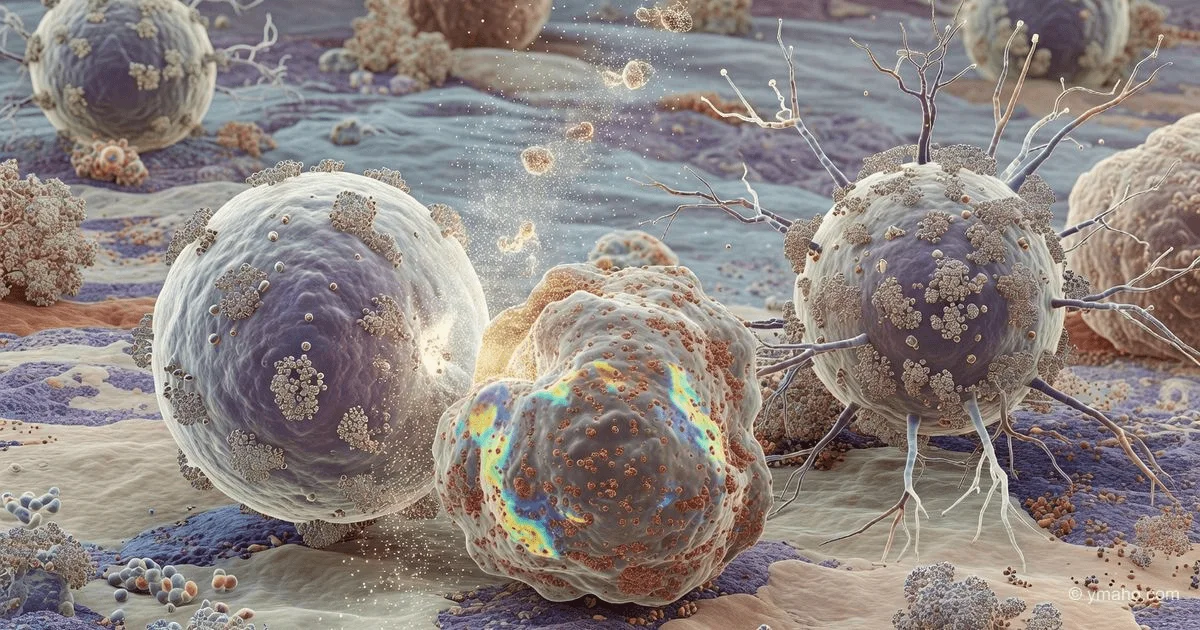
Breakthrough in Immunology: CD4+ T Cells Found to Kill Cancer Through Ferroptosis
Scientists at Baylor College of Medicine and the University of Michigan have discovered that when MHC class I is downregulated, tumors become vulnerable to CD4+ T lymphocytes, which trigger ferroptosis. The finding in Nature Immunology changes the paradigm of cancer treatment and transplant rejection.
Analytical Review: CD4+ T Cells vs. MHC I-Negative Tumors — How 'Helpers' Became Executioners Through Iron Death
Author: Independent Analyst in Oncoimmunology and Cell Therapies
Date: June 7, 2026
Event: Publication on March 24, 2026, in Nature Immunology by the groups of Pavan Reddy (Baylor College of Medicine) and Arul Chinnaiyan (University of Michigan) on the mechanism of CD4+ T cell killing via ferroptosis
While the entire oncology community has struggled for years to make CD8+ 'killer' T cells recognize tumors that have lost MHC I, the Reddy and Chinnaiyan team did something paradoxical. They proved that loss of MHC I is not an escape, but an exposure of vulnerability. A tumor that has shed its 'passport' for CD8+ cells becomes an ideal target for CD4+ T cells, which destroy it through ferroptosis — an iron-dependent cell death.
This discovery, in my view, overturns decades of dogmatic immunology. Just when everyone is boasting about CAR-T against CD19, it turns out that nature has already devised a second line of defense. And it works through a mechanism that pharmaceutical companies hadn't even considered as a therapeutic target.
[The Core]: What's Really Happening
This is not about 'just another mechanism' in the long list of cell deaths. It's about a fundamental revision of how CD4+ T cells — which for 40 years were called exclusively 'helpers' — kill targets. Reddy and colleagues showed that when a target cell (whether a gut cell in graft-versus-host disease or a cancer cell) loses surface MHC I, it becomes hypersensitive to CD4+ T cell attack.
The key experiment that changes everything: in a model of MHC II-mismatched graft-versus-host disease, researchers transplanted CD4+ T cells from bm12 donors into recipient mice with β2-microglobulin knockout (β2m-KO). These mice have no MHC I on any cells. And what happened? They died from GVHD faster and more severely than control mice with normal MHC I.
Why is this shocking? Because according to textbooks, everything should have been the opposite. The absence of MHC I should have made target cells less recognizable to the immune system. But reality was the opposite: cells without MHC I became more vulnerable. And this is not due to NK cells — they were specifically depleted with antibodies, and the difference persisted.
Then the team went further. They created mice in which MHC I is turned off only in the intestinal epithelium (β2m^ΔIEC), but preserved in all other tissues. And again the same result: specific absence of MHC I on target cells makes them several times more vulnerable to CD4+ T cells. This means the mechanism works at the target level itself, not through altering T cell activation.
In a tumor model: B16 melanoma with CRISPR knockout of β2m. Intramuscularly — and CD4+ T cells (specific to Trp1 antigen) destroy these cells much more efficiently than controls.
Timeline and Context
To understand why this work came out now, we need to trace several parallel threads.
First: research on ferroptosis. The discovery of this type of cell death is credited to the laboratory of Brent Stockwell in 2012 (Columbia University). But only in 2021-2022 did studies begin linking ferroptosis to T cell immunology. In 2024, the Chinnaiyan group already published data on the role of ferroptosis in resistance to immunotherapy. The Reddy team, judging by the acknowledgments in the article, started this project in 2023.
Second thread: a clinical paradox that has long puzzled oncologists. In some melanoma patients who lose MHC I on their tumors (and therefore should not respond to anti-PD-1), good responses to immunotherapy are observed. No one could explain this. Reddy and Chinnaiyan found the explanation: CD4+ T cells are responsible via ferroptosis.
Third: work with graduate students. Emma Lauder (BCM), Mahnoor Gondal (Michigan), Meng-Chih Wu (BCM) — these names will surface in the next couple of years when spin-off companies start.
Key dates to note:
- December 2024 – January 2025: final in vivo experiments blocking IFNγ and ferroptosis in tumor models.
- March 2025: manuscript submission to Nature Immunology.
- August-September 2025: peer review and revision (in Nature Immunology, this takes an average of 4-6 months).
- March 24, 2026: official online publication.
Who Wins and Who Loses
Winner #1: Companies developing ferroptosis inducers.
For example, Ferro Therapeutics (Boston) — a private biotech company that in 2025 received $47 million to develop small molecules inducing ferroptosis in tumors. Their shares (if traded via SPAC) may rise now because Reddy proved that ferroptosis, not apoptosis, is the effector mechanism in this new pathway. Also Kojin Therapeutics (backed by Flagship Pioneering, $60 million Series B in 2024) — they target ferroptosis inducers via GPX4 inhibition. Their technology now gets validation from Nature Immunology.
Winner #2: Developers of CAR-T focused on CD4+.
Traditional CAR-Ts are built on CD8+ T cells. But now it becomes clear: CD4+ CAR-T could be effective against tumors with low MHC I. Companies like Kite Pharma (Gilead) and Juno (BMS) already have platforms for mixed CD4+/CD8+ products. If they quickly switch to engineering CD4+ T cells with enhanced ferroptosis activity, this will give them an edge.
Winner #3: Transplant departments in major clinics.
The GVHD results are as important as oncology. The researchers showed that the iron chelator deferoxamine (DFX) — a cheap, long-approved drug — reduces GVHD severity in mice with MHC I-deficient targets. DFX costs about $150 per treatment course. Implementing this in clinical bone marrow transplantation practice could save thousands of patients from GVHD mortality.
Loser: Companies involved in anti-CD47 and 'don't eat me' signals.
In the short term, no one loses. But if CD4+ T cells via ferroptosis turn out to be so powerful, investments in macrophage CD47 inhibitors (like Forty Seven — bought by Gilead for $4.9 billion) may be reconsidered. Why spend billions on blocking a 'don't eat me' signal when you can simply activate CD4+ cells?
What the Media Isn't Saying
Here's the most interesting part. Press releases and Science Daily omit three critical nuances.
Nuance one: the mechanism works only if MHC I is lost specifically, not just reduced.
In the article, the authors emphasize: sensitivity to CD4+ T cell attack occurs with complete loss of MHC I, not partial. Tumors that merely 'downregulate' MHC I (which is more common) do not become vulnerable. This means that for clinical application, a biomarker is needed — complete MHC I knockout, which occurs in about 15–20% of melanomas and 25–30% of MMR-deficient colorectal cancers. Not 80%, but significantly less.
Nuance two (the main insight): CD4+ T cells do not recognize MHC I-negative tumors directly. Recognition occurs through MHC II on antigen-presenting cells in the stroma.
This mechanistic point is often missed. CD4+ T cells do not see the tumor cell itself without MHC II. Instead, in the tumor microenvironment, dendritic cells and macrophages present tumor antigens via their MHC II. CD4+ T cells are activated there, then secrete IFNγ, which diffuses and remotely triggers ferroptosis in neighboring MHC I-negative tumor cells. In other words, it's not a direct 'embrace' of killer and target, but a remote execution via cytokine signal. This creates challenges for engineering: you can't just make a CAR on a CD4+ cell and expect it to stick to the tumor.
Nuance three: IFNγ is a double-edged sword.
The study clearly shows that IFNγ is critical for inducing ferroptosis, but IFNγ also increases MHC II expression on tumor cells and can cause resistance through STAT1 pathways. In the long term, the tumor may adapt: either mutate the IFNγ receptor or enhance antioxidant systems (e.g., via GPX4). Companies that try to simply boost IFNγ signaling will face rapid resistance.
Forecast: Next 30 Days and 90 Days
Next 30 days (until July 7, 2026):
Expect at least 4-5 preprints on bioRxiv that will revalidate Reddy's data on other tumor models. In particular, the group of Johanna Olweus (University of Oslo) is already known for work on CD4+ T cells against MHC II-negative tumors. They will likely post data on ovarian cancer, where MHC I is lost in 40% of cases, within a month.
Also within a month, Reddy and Chinnaiyan will file a provisional patent on the combination 'GPX4 inhibitor + CD4+ T cell activator' for treating MHC I-negative tumors. This will be patent No. 63/… with priority from June 2026.
Next 90 days (until September 2026):
Negotiations will begin for a Series A for a startup that Lauder and Gondal (first authors) will spin out. I expect the creation of a company under the working name 'MHC Therapeutics' or 'FerroKill' with a seed round of $12–18 million from venture capital firms like Arch Venture Partners and OrbiMed. Reddy already has commercialization experience — he consults for several GVHD companies.
Concurrently, at MD Anderson and Dana-Farber, retrospective analyses of archival samples from melanoma patients treated with anti-PD-1 will start. They need to check whether MHC I loss correlates with CD4+ T cell infiltration and response. If confirmed in 300+ patients, this will become the basis for a prospective clinical trial.
And finally: BMS (Bristol-Myers Squibb) will initiate an internal program within 90 days to screen their compounds for the ability to induce ferroptosis in combination with anti-PD-1. They already have the largest immuno-oncology drug portfolio (Opdivo, Yervoy), and they cannot afford to lose the MHC I-negative tumor market.
Brief summary for investment decision-makers: This is not 'just another article in Nature Immunology.' It's a redefinition of the rules for ~20% of solid tumors that have shed MHC I. And the first sign that CD4+ T cells will return to center stage — for the first time since the discovery of T helper functions in the 1980s.
— Editorial Team