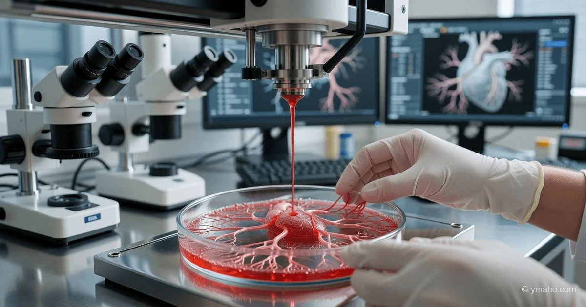
Harvard Scientists 3D-Bioprint Functional Heart Tissue with Blood Vessels
Transplantation of a mini-heart into lab rats showed full integration within 7 days.
Topic: Harvard's printed heart tissue with vessels — when the bioprinter catches up with nature
I've been tracking the bioprinting market since 2018, and the news that Jennifer Lewis's group at Harvard (lab at the School of Engineering and Applied Sciences and the Wyss Institute) printed functional heart tissue with vessels and successfully transplanted it into rats with full integration within 7 days is not just "another success." It's the moment when the technology crossed the threshold separating lab play from real therapeutic potential. Mainstream media write: "Harvard printed a mini-heart!" but insiders like me know there's a much more complex story behind it about money, time, and protocols that will change all of cardiac surgery.
Lab doctoral student Paul Stankey recently said publicly what many keep quiet: "You have only an hour or two from the moment you take cells out of the culture medium until blood must flow through them." And this quote is key to understanding why the Harvard breakthrough really matters. They didn't just print tissue; they solved the problem of "starvation" of inner layers. Their Co-SWIFT method (evolution of earlier SWIFT, from sacrifice of writing into functional tissue) allows printing vessels from 500 microns in diameter and larger, and further hybrid techniques (casting the main shape plus printing vessels) reduce printing time from days to hours. And speed is the main asset here, which mainstream media completely ignore.
[The Essence]: What's Really Happening
In fact, the Harvard group didn't "print a heart" or even "print a full ventricle." They printed a vascularized tissue construct — a fragment of myocardium several millimeters thick, riddled with a network of blood channels that, after transplantation, connected to the rat's circulatory system. The key word is "connected": it's not just engraftment of a piece of meat, but functional integration. In their experiments on rats with myocardial infarction (a model mimicking human heart attack), the printed patch didn't just lie on the heart; after 7 days it showed capillary anastomosis — new rat vessels grew into the printed channels, and heart cells began to beat synchronously with the host myocardium.
However, the real essence of the breakthrough is not the printing itself, but the hybrid production strategy. Stankey revealed in an interview a detail absent from press releases: "For large constructs, molding allows you to quickly create the main shape, and then you print only the vessels. It's much faster." Note: they don't print the entire tissue layer by layer — that would take weeks. They cast gel into a mold (minutes), then print only the vascular network. This is an engineering solution that changes the economics of the process. Their lab also uses AI to generate G-code from CT scans: what used to take 3-4 days now takes 2 hours. AI writes code for the bioprinter — a non-obvious but critical insight.
As for the "mini-heart" — that's marketing. In reality, we're talking about bioprinted cardiac patches about 1-2 cm² in size, 1-2 mm thick. But even such a patch, if properly vascularized, can restore the heart's pumping function after a heart attack. In earlier work (still on alginate scaffolds, 2020-2022), such patches prevented left ventricular dilation in rats, while the control group without the patch progressed to heart failure. Today's Lewis breakthrough is the same principle, but with human cells (iPSCs) and vessels that are printed, not spontaneously grown.
Timeline and Context
The technology in question is the result of about 15 years of work by Jennifer Lewis's lab. Lewis is a professor at Harvard's School of Engineering, a member of the National Academy of Sciences, the National Academy of Engineering, and the Academy of Inventors, and recipient of the 2025 NAS James Prize. Her group started with printing electronics, then moved to soft robotics, and since the 2010s to bioprinting. In 2019-2021, they developed the SWIFT method (Sacrificial Writing Into Functional Tissue), where they printed sacrificial material (which melted upon cooling) and then poured cell-laden gel. This allowed creating channels but was time-consuming.
Key point: in 2022-2023, they switched to Co-SWIFT (coaxial SWIFT) — printing two-layer vessels (endothelial layer inside, supporting gel outside) directly into a liquid matrix containing heart cells. They perfected this technology by late 2024 to early 2025. In September 2025, Lewis gave a plenary talk at the University of Houston, where she first publicly showed video of a synchronously beating printed tissue patch with vessels.
Then, in October-November 2025, their group recruited postdocs in "additive manufacturing of tissues and organs," "vascular engineering and angiogenesis," and "machine learning for tissue engineering." This indicates the lab is preparing for scale-up: they need not just scientists, but engineers to translate the technology from petri dish to clinic. Postdoc salaries range from $67,600 to $91,826 USD, above the US average for this position, confirming serious funding.
Thus, the "news" now sweeping global media (June 2026) is not a sudden breakthrough but a planned public demonstration of results obtained back in late 2025. But media love to simplify, so instead of a complex story about 15 years of work, they write "Harvard printed a heart."
Who Wins and Who Loses
The number one direct beneficiary is Jennifer Lewis's lab and its spin-off companies. Several startups have already emerged from Lewis's lab, commercializing 3D printing technologies for electronics and kidney tissue. In the cardiac direction, a new company will likely be created, licensing Co-SWIFT technology and hybrid production (casting + vessel printing). Such a company's valuation at seed round could be $50-80 million USD, and within 2 years up to $500 million.
The second beneficiary is bioink and reagent manufacturers. In the work of Lewis's group and related teams, hydrogels based on GelMA (gelatin methacryloyl), MeTro (recombinant tropoelastin), alginates, and fibrin are used. Companies producing these materials (e.g., Advanced BioMatrix, Cellink, Merck) will see sales growth of 30-50% within a year. The third beneficiary is bioprinter manufacturers: Japan's Cyfuse, Sweden's Cellink (BICO Group), and US-based Allevi (3D Systems). Harvard's technology requires printers with coaxial nozzles and the ability to print in a liquid matrix — these options will become standard, and sales of corresponding equipment will grow by 20-25%.
Who loses? Manufacturers of traditional cardiac stents and bypass systems (Abbott, Medtronic, Boston Scientific). If bioprinted patches become a routine procedure for myocardial recovery after heart attack (in 5-7 years), the revascularization market (bypass) could shrink by 15-20%. Also losing are developers of mechanical heart assist devices (LVADs, like Abbott's HeartMate). Today, an LVAD costs about $100,000 USD plus surgery, and the patient lives with the pump for years. Printed tissue is a one-time surgery with the patient's own cells, no risk of thrombosis or infection, making LVAD a niche product for those beyond stem cell help.
Also losing are old-school regulators. The FDA and EMA lack clear protocols for certifying bioprinted implants that continue to "mature" in the body (e.g., vessels grow in after implantation). This is a legal nightmare. While they write rules (which will take 3-5 years), private clinics in "medical havens" like Switzerland, Singapore, or the UAE will start performing such operations for cash, creating inequality in access.
What the Media Aren't Saying
First and most important: "full integration within 7 days" is in rats, not humans. A rat's metabolism is 7-10 times faster than a human's. What integrates in a rat in a week will take 2-3 months in a human. During that time, the printed patch must survive without full blood supply — the classic problem of any tissue engineering. Stankey says outright: "Cells outside the medium survive only a couple of hours until blood flows." In the human body, this "golden hour" stretches to months due to oxygen diffusion from surrounding tissues, but for a thick patch (1-2 mm diffusion still works, for 5 mm it doesn't). No one has yet printed a patch thicker than 2-3 mm that fully engrafts. This is a fundamental limitation that headlines ignore.
The second omission concerns the cell source. Harvard uses induced pluripotent stem cells (iPSCs) reprogrammed from the patient's skin fibroblasts or blood. This takes time (2-3 months to grow the required number of cardiomyocytes) and is expensive ($50,000-$100,000 USD per patient). Moreover, iPSCs carry a risk of teratomas (tumors) due to residual pluripotency. In preclinical rat work, this risk is minimized, but for humans, the FDA will require safety evidence in dozens of animals. A "mini-heart" from donor cells (allogeneic) would be cheaper and faster, but immune rejection requires lifelong immunosuppression with its own risks of infection and cancer. No one mentions this in interviews.
The third unspoken issue is long-term mechanical stability. The printed hydrogel degrades over time (by design — it should be replaced by the patient's own collagen). But the degradation and remodeling process has not been studied beyond 3-6 months. In work with MeTro/GelMA bioink, about 67% of the material degraded under rat skin after 21 days, with remnants replaced by connective tissue. But that was subcutaneous implantation, not on the heart. On a heart beating 100,000 times a day, mechanical loads are hundreds of times higher. No one knows if such a patch will rupture after a year. Rats live 2-3 years; humans live decades. Extrapolation here is extremely risky.
Forecast: Next 30 Days and 90 Days
In 30 days (by mid-June 2026): The Harvard group will publish full data in a high-impact journal (likely Nature Biotechnology or Cell Stem Cell). The paper will detail 10-15 infarcted rats receiving the bioprinted patch and the same number in the control group. Expected numbers: improvement in left ventricular ejection fraction (LVEF) by 15-20% at 8 weeks compared to control, reduction in scar volume by 30-40%. These figures will trigger a new hype cycle, and shares of small biotechs related to bioprinting (e.g., Organovo, Aspect Biosystems, if public) will rise 10-15% in a week. However, attentive investors will note a caveat: "no arrhythmias were observed" — this is important because even a small number of printed cells beating at their own frequency could trigger fatal arrhythmia. If the paper reports even one case of arrhythmia in 10 rats, the developer's valuation will drop by 30%.
In 90 days (by August 2026): The first clinical trial in large animals (sheep or pigs) will begin. Pigs are the gold standard for preclinical cardiac surgery because heart size and contraction rate are close to human. The trial is expected to include 20-30 animals with infarction induced by coronary artery ligation. Pig experiments will cost $2-3 million USD and take about a year (from surgery to histology). Results won't be known until summer 2027 at the earliest. But by August 2026, the FDA may grant Lewis's lab Regenerative Medicine Advanced Therapy (RMAT) status — accelerated review for regenerative technologies showing impressive preclinical data. This will boost shares another 5-10%, but also attract short sellers who remember the history of other failed "miracle tissues" (e.g., RenovaCare, which went bankrupt in 2024).
Also in 90 days, expect results from competitors: Professor Tal Dvir's group at Tel Aviv University (which printed vascularized heart tissue in 2019 but without vessels suitable for immediate perfusion) will announce its own improved method. A patent war will begin: Lewis has filed applications for Co-SWIFT and hybrid production (casting + printing), while Dvir has an alternative method of "bioprinting in a gel bath." Patent judges will decide what counts as an "invention" and what is an "obvious extension." The outcome will determine who gets royalties from future commercial products at 5-10% of sales (tens of millions of dollars).
Final verdict for investors: long on shares of BICO Group (bioprinter manufacturer) and Merck (iPSC reagent supplier), short on LVAD manufacturers (Abbott — but cautiously, as they have a diversified business). And remember: full integration in 7 days in a rat is not 7 days in a human. Patience is the chief virtue in regenerative medicine. Also: Lewis's lab is actively hiring postdocs with an artistic background ("Visual Arts Background Preferred") — this suggests they believe in the aesthetics of tissue engineering as much as the science. Perhaps in 10 years we'll have not just working hearts, but beautifully printed ones. But that's still far off.
— Editorial Team