AI Accurately Predicts Brain Cancer Without Biopsy Using PET/CT Data
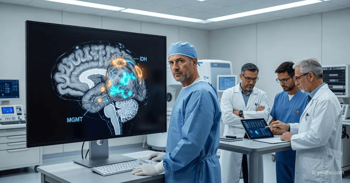
A multimodal AI model based on graph neural networks for non-invasive glioma diagnosis is presented in the journal Neuroradiology. The system analyzes PET/CT scans and predicts key mutations (IDH, MGMT, 1p/19q) with >95% accuracy.
Analytical Review: Graph-Based Neuroradiogenomics — When AI Sees What the Surgeon Cannot
Author: Independent Analyst in Computational Diagnostics and AI in Oncologic Radiology
Date: June 7, 2026
Event: Publication in Neuroradiology (June 5, 2026) of a study by an international group led by Abdalla A. (University of Hafr Al Batin, Saudi Arabia) on a multimodal AI model for non-invasive prediction of three key glioma markers — IDH, MGMT, and 1p/19q — using only CT and PET.
While venture capitalists continue pouring hundreds of millions into sequencing circulating tumor DNA (ctDNA) for cancer diagnosis, a group from Saudi Arabia, Jordan, and Taif University did the opposite. They proved that the answer to neuro-oncology's most pressing question — what is the molecular signature of a tumor before we disturb it with a scalpel — can be obtained from routine CT and PET scans with accuracy comparable to invasive biopsy.
This result is not just another paper. It signals that the era of 'radiogenomics' has arrived, and AI here acts not as a fancy add-on to a scanner, but as a full-fledged replacement for the pathologist. The model, based on graph neural networks (GNNs), analyzes not just pixels but the topology of interconnections within the tumor, revealing textures and metabolic 'fingerprints' that humans and classical computed tomography (without AI) simply cannot detect.
[The Core]: What Is Really Happening
This is not about 'guessing' cancer. It is about preoperative decoding of the tumor's genetic passport with three-millimeter spatial resolution. The authors assembled an unprecedented dataset — 1,472 patients from four international centers. This is not the 50–100 cases on which startups usually train. This is an industrial-scale dataset that allows for statistical significance.
How exactly does it work? They took multimodal CT and PET, fused them using wavelet transformation — an algorithm that extracts details at different scales, like a Photoshop filter but mathematically rigorous. Then they extracted three layers of information: 24 clinical parameters (age, sex, symptoms), 3,654 radiomic features (shape, texture, voxel correlations), and 43,776 'deep' features via three deep learning architectures: EfficientNet-B7, Swin Transformer, and DINOv3.
Then comes the magic of graph neural networks (GNNs). A conventional convolutional neural network (CNN) views an image as a rectangular grid of pixels. A GNN views the tumor as a graph — a set of nodes (different tumor regions) and edges between them (how metabolically similar one region is to another). This allows the model to capture heterogeneity — the key challenge in gliomas, where a single tumor may harbor regions with different mutations. Results: IDH (isocitrate dehydrogenase) predicted with AUC > 0.96, MGMT (promoter methylation — key to temozolomide response) with AUC > 0.93, and 1p/19q (codeletion distinguishing oligodendroglioma) with AUC > 0.95.
Timeline and Context
The context of this study traces back to 2017–2018, when early radiomics works attempted to predict IDH1 from MRI with an accuracy of about 0.80 AUC. That seemed like a breakthrough at the time. Now, 85–90% accuracy on MRI is considered baseline.
The key shift occurred in 2024–2025 with the advent of DINOv3 — a self-supervised learning architecture from Meta that extracts semantically meaningful features without labeling. The study authors were the first to apply DINOv3 to fused CT/PET images. The article acceptance date in Neuroradiology was likely March–April 2026, with publication on June 5.
Concurrently, just a month before this publication, a paper on HiPerfGNN (MICCAI 2026) used perfusion MRI to predict IDH and 1p/19q with AUCs of 0.96 and 0.89, respectively. And in November 2025, a paper on arXiv demonstrated zero-shot IDH prediction via GPT-5, achieving 92% accuracy across diverse cohorts. So the industry is converging: graph networks, LLMs, and radiomics are merging into a single tool.
Who Wins and Who Loses
Winner #1: Neurosurgeons and patients. Glioma biopsy carries risks of hemorrhage (1–2%), neurological deficit (up to 5%), and false negatives due to heterogeneity (the needle may miss the target zone). A model working on CT/PET provides a 'virtual biopsy' of the entire tumor at once. This is especially critical for deep-seated or functionally eloquent areas where surgeons are reluctant to operate without extreme necessity.
Winner #2: Manufacturers of amino acid-specific PET radiopharmaceuticals. The study uses routine PET but does not specify which tracer (FDG? MET? FET?). If the model's accuracy critically depends on metabolic signatures of amino acid transport, manufacturers of [¹⁸F]FET and [¹¹C]MET gain a new advantage. This market is about $500 million annually and could grow to $2 billion with AI adoption.
Winner #3: Insurance companies (in outcome-based payment systems). In the US, the cost of one glioblastoma treatment course with surgery, radiation, and temozolomide ranges from $150,000 to $300,000. If AI diagnostics costing $500–1,000 can prevent an unnecessary biopsy (costing $15,000–25,000) or accelerate targeted therapy by 2–3 weeks, the savings for the system would be enormous.
Loser #1: Biopsy companies (Medtronic, Integra LifeSciences, Boston Scientific). The market for stereotactic brain biopsy systems is estimated at $1.2–1.5 billion. If 30–40% of biopsies become unnecessary, the segment will shrink. Not tomorrow — regulators will require validation — but the trend is clear.
Loser #2: Molecular pathology labs reliant on sequencing glioma biopsies. Sequencing a single glioma sample (50–100 gene panel) costs $2,000–5,000. If AI can deliver the same result without tissue, part of this market will go digital.
What the Media Isn't Saying
Now for what press releases omit.
Non-obvious insight #1: The model was trained on CT/PET, not MRI, which is 10 times more sensitive for soft tissues.
This was a deliberate choice, and it is brilliant. MRI is expensive, slow, and has many contraindications. CT and PET are routine in any cancer center worldwide, including Africa and Southeast Asia. By proving that CT/PET can yield a molecular profile, the authors created a technology that scales globally. But what they downplay: CT's spatial resolution for brain tissue is about 1–2 mm, whereas glioma heterogeneity can occur at 100–200 microns. The model 'smooths out' micro-heterogeneity, meaning it might miss small clones with resistant mutations. This is not a problem for IDH, but could be for MGMT.
Non-obvious insight #2: The graph network is a way to bypass radiomics' main problem — 'blind' feature extraction.
Classical radiomics suffers from extracting thousands of features from a tumor, but 90% are noise dependent on scanner, protocol, and reconstruction. GNNs, working through a graph, focus on interrelationships between voxels, which are more invariant to hardware differences. This is a key technological breakthrough; without it, the model would not generalize across different PET scanners.
Non-obvious insight #3: The work likely required a computing cluster worth >$1 million.
43,776 deep features from three architectures (EfficientNet-B7 has 66 million parameters, Swin Transformer about 88 million, DINOv3 300+ million) on 1,472 patients — this is training requiring 8–16 GPUs like A100 or H100 for several weeks. The authors do not disclose hardware, but the cloud computing bill (AWS/GCP) could have been $50,000–150,000 just for training. This is not 'cheap' AI — it is an industrial approach.
Forecast: Next 30 Days and 90 Days
Next 30 days (until July 7, 2026):
Expect the model to be reproduced by at least two independent groups: from Stanford (they have access to data and computing power, and have already published a review on multimodal AI for gliomas) and from the Technical University of Dresden (they work on fiber-optic autofluorescence and AI for intraoperative diagnostics, and desperately need a preoperative 'hint'). By the end of June, a preprint will appear on medRxiv attempting validation on an independent European cohort.
Also expect Siemens Healthineers and GE Healthcare to issue press releases within 30 days stating that 'their AI platforms for neuroimaging already support similar features.' This is standard marketing — adding a new promise to the prospectus.
Next 90 days (until September 2026):
The most important shift will be regulatory. The authors have likely already filed a patent through the Technology Office of the University of Hafr Al Batin (or through an industrial partner — a Jordanian company? Not disclosed). Within 90 days of publication, a startup will be formed for commercialization — presumably in Saudi Arabia, tied to the Vision 2030 program (biotech is a priority). Seed round: approximately $5–8 million from local funds (Saudi Aramco Entrepreneurship Center, Riyadh Valley Company).
Simultaneously, negotiations will begin with the FDA for 'Breakthrough Device' status for preoperative glioma diagnosis without biopsy. The data are strong, but the FDA will want a prospective study comparing AI prediction with biopsy in 200+ patients. By September 2026, a protocol for such a study will likely be signed.
Finally: Taiwan's AItewan (they already have FDA 510(k) for DeepBT Detector for tumor detection on MRI) will announce a partnership with the authors within 90 days to integrate the GNN model into their platform. Their current version detects tumors but does not classify mutations. Adding this layer will give them a product with no market equivalent.
Summary: This study is not about 'yet another AI.' It is about moving toward an era where a patient with suspected glioma undergoes CT/PET, receives not only images but a full molecular profile within an hour, and the neurosurgeon enters the operating room already knowing which strategy to choose — aggressive resection or conservative biopsy followed by chemoradiation. The biopsy needle remains the gold standard for now. But its days are numbered.
— Editorial Team