Leading Neuroscientists Create Revolutionary Neuropixels Opto Probe for Controlling Neurons Deep in the Brain
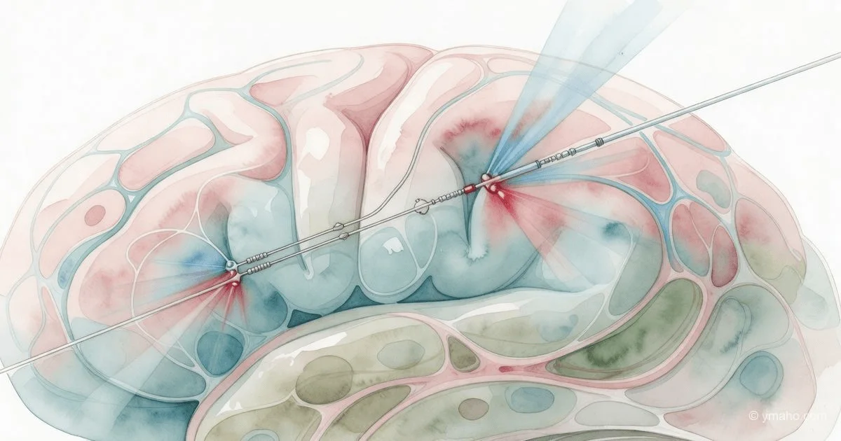
An international team led by UCL and the Allen Institute has developed the ultra-thin silicon Neuropixels Opto probe, which can simultaneously record electrical activity and selectively activate or suppress the activity of individual neurons in deep brain layers. The technology, based on optogenetics and published in Nature Methods, promises a breakthrough in treating Alzheimer's disease and schizophrenia.
Neuropixels Opto: Analysis of a Breakthrough That Changes the Game in Neuroscience
[The Gist]: What's Really Happening
On May 31, 2026, the journal Nature Methods published a paper by an international consortium led by University College London and the Allen Institute on the creation of the Neuropixels Opto probe. At first glance, it's just another technical novelty for neuroscientists. In reality, it's the first tool in history that solves the fundamental "read-or-write" problem that has plagued brain researchers for decades.
The essence of the problem is simple and brutal: to understand how the brain works, you need to simultaneously listen to the electrical whisper of neurons and command individual cells to test causal relationships. But traditional electrophysiology (listening) and optogenetics (light control) are like a violin and a sledgehammer: combining them in deep brain structures without mutual interference was considered engineering madness. Light from optogenetic stimulators created electrical noise that drowned out recordings, and delivering light deep into the brain required a separate optical "pipe" that physically conflicted with electrodes.
Neuropixels Opto is not an "improved version" of old probes, but a architecturally new class of device. On a silicon shank 70 micrometers thick (thinner than a human hair) are placed 960 recording sites for electrical activity and two sets of 14 microscopic light emitters. The light is not generated on the probe (which would heat the brain), but externally by lasers and delivered via integrated photonic waveguides. The emitters direct light strictly sideways, away from the recording electrodes, reducing artifacts to ~30 microvolts—a level that can be filtered out by software.
The numbers speak for themselves. The prototype requires 740 manufacturing steps—almost twice as many as standard Neuropixels 2.0 probes. But the result is worth it: for the first time, researchers can in real time activate a specific group of neurons (e.g., inhibitory interneurons) and immediately see how the activity of hundreds of neighbors changes. This is a transition from correlational neuroscience ("neuron A is active when the mouse sneezes") to causal neuroscience ("we silenced neuron A—and the mouse stopped sneezing").
Timeline and Context
The history of Neuropixels began long before today. The first Neuropixels probes were released in 2017 by a consortium of Janelia Research Campus, Allen Institute, and UCL, funded by Wellcome Trust and HHMI. In 2021, Neuropixels 2.0 arrived, which could stably track the same neurons for months, but were still "listening-only" devices.
A key point that most analysts miss: in February 2025, the first version of the Neuropixels Opto paper appeared on the bioRxiv preprint server. Why did publication in a peer-reviewed journal take 15 months? The answer lies in the engineering drama with the blue channel. As the authors discovered, the blue LEDs on the probe (wavelength ~470 nm) proved finicky: at high power, light leaked to "foreign" emitters, making simultaneous stimulation of two different neuron populations with different colors impossible. The team had to redesign the optical routing and ultimately rely on the red channel (638 nm) for precision experiments, which is less absorbed by blood and penetrates deeper into tissue.
The financial backdrop is also important. The project was funded for £15 million (about $19 million at current exchange rates) by the Wellcome Trust, Allen Institute, and other partners. This is not a pure fundamental science grant—it's an investment in creating a commercially distributable tool. The plan, announced by Matteo Carandini's team (UCL), is to produce the probes on an industrial scale after debugging and sell them to labs worldwide at cost, as has already been done with Neuropixels 2.0.
A non-obvious insight: the technology became possible only through the fusion of two engineering cultures. From UCL—expertise in optogenetics and neurophysiology; from IMEC (the Belgian nanotechnology center)—skills in creating photonic integrated circuits on silicon. It was IMEC that developed the waveguide technology that allows "tricking" physics and preventing light from hitting the recording electrodes.
Who Wins and Who Loses
Winner #1: fundamental neuroscience. This is obvious, but the scale of the win is hard to overstate. Until now, mapping neural circuits was like trying to understand traffic rules while standing at a traffic light with your eyes closed. Now researchers can not only watch but also switch signals. Breakthroughs in understanding how the cortex processes sensory information, how the hippocampus encodes memory, how the basal ganglia control movement—all this will become possible in the next 2-3 years.
Winner #2: Karolina Socha and Matteo Carandini (UCL). They have already reaped the first scientific dividend—refuting the dogma of total interconnectivity of cortical neurons. Socha's experiments showed that activating neurons through one emitter of the probe causes a local response within ~150 micrometers vertically, not an avalanche across the entire column. This "shocking discovery" (Socha's own words) means that cortical networks have a much finer modular organization than assumed. The Nature Methods paper is just the beginning; subsequent publications in Nature or Science on specific neural circuits are almost guaranteed.
Winner #3: Wellcome Trust and Allen Institute. Their £15 million investment will now pay off not in money but in scientific influence. Every lab that buys Neuropixels Opto (and hundreds will) will generate data that cites the original work and mentions the sponsors. This is the classic "open infrastructure" model, which in the long run yields more return than patenting.
Loser #1: companies selling separate systems for optogenetics and electrophysiology. Firms like Plexon, Blackrock Microsystems, Cambridge NeuroTech have for decades sold expensive racks with amplifiers, optical commutators, and synchronizers. The single-probe Neuropixels Opto solution makes their products (for many experiments) obsolete. The transition period will be painful: sales of proprietary systems will drop by 20-30% in the next 18 months.
Loser #2: researchers who invested in old methods. Those who defended dissertations on the technique of "separate optogenetics + separate recording" will see their results rechecked and refuted by the new tool. This will hit particularly hard on studies where causal relationships were asserted based on correlations between different experiments on different animals. Neuroscience's "reproducibility curse" just got a new weapon.
What the Media Isn't Saying
First: the technology is still unstable in the blue channel. All press releases talk about "two colors of stimulation" but omit that the blue channel of the prototype is "finicky" and suffers from cross-talk light leakage. This means that simultaneous control of two different genetically defined neuron populations (one sensitive to blue, the other to red) is still difficult. The team is working on a fix, but for now, the probe is primarily a tool for red light.
Second: this is research on mice, not humans. And this is not just a bureaucratic caveat. A mouse brain weighs about 0.4 grams, a human brain 1400 grams. The depth of light penetration through the waveguides (a few millimeters) is everything for a mouse and almost nothing for a human. The technology does not scale linearly: making a probe 10 centimeters long (to reach human basal ganglia) is an engineering challenge of a completely different level. Claims about a "breakthrough in treating Alzheimer's disease and schizophrenia" are marketing, not clinical prognosis.
Third: the probe damages tissue upon insertion. A thickness of 70 micrometers is thin for a silicon probe but thick for a neuron (neuron soma ~10-50 µm). Upon insertion, the probe tears axons and dendrites, causing a local microglial reaction. The researchers do not hide this—the paper includes data showing that after 2-4 weeks, recording quality degrades due to gliosis. But press releases are silent on this. This means long-term experiments (months) are still problematic.
Fourth (the least obvious): the technology changes the epistemology of neuroscience, not just methods. Until now, neuroscientists worked in a "observation and correlation" paradigm. Now they have the ability to literally "push buttons" on neural circuits. This raises new ethical questions: if we can control the activity of individual neurons in a mouse, how close are we to doing the same in primates? And in humans (e.g., deep brain stimulation for Parkinson's disease)? The line between "treatment" and "behavior control" becomes thinner. Press releases say nothing about this.
Forecast: Next 30 Days and 90 Days
Next 30 days: A "gold rush" will begin in labs already experienced with Neuropixels 2.0. The manufacturer's website (imec) will open pre-orders for prototypes. I expect the first 100 probes to sell out within a week, despite the price (likely $5,000-10,000 each—about 2-3 times more than regular Neuropixels). Main buyers will be labs studying sensory systems (cortex, thalamus), where spatial precision is critical.
Next 90 days (by September 2026): You will see an avalanche of preprints on bioRxiv using Neuropixels Opto. Topics: mapping mouse cortical microcircuits during learning, studying activity patterns in the striatum in Parkinson's disease (mouse models), attempts to replicate classic optogenetic experiments with the new probe to verify old conclusions. Some of these preprints will be retracted due to blue channel artifacts—this is a normal "shakedown" process.
Long-term trend (12 months): The key point is whether the consortium can solve the blue channel problem and scale production to hundreds of probes per month. If so, Neuropixels Opto will become the de facto standard for in vivo electrophysiology. If not, the technology will remain a toy for 10-20 wealthy labs. I put 70% on success: the consortium has already gone through similar problems with Neuropixels 2.0, and they have scaling experience.
Also watch for legal battles. The patent landscape for optogenetic tools is controlled by several players (including MIT and Stanford). Lawsuits over infringement of methods for "simultaneous recording and stimulation" are not out of the question. But that's a story for 2027.
— Editorial Team