AI MouseMapper: A New Platform for 3D Mapping of Organ Damage in Obesity
German scientists (Helmholtz Munich) have created an AI platform called MouseMapper for whole-body analysis. The AI revealed damage to the trigeminal nerve in mice and humans due to obesity, explaining the reduced sensitivity in patients.
Analytical Review: MouseMapper — When AI First Saw the Body as a Whole
Author: Independent analyst in AI for biomedical research and systems biology
Date: June 7, 2026
Event: Publication in Nature on May 19, 2026, of a study by the group of Ali Ertürk (Helmholtz Munich) and Maximilian Schefer (LMU Munich) on the MouseMapper platform — the first AI tool for holistic 3D whole-body analysis at cellular resolution.
While the medical AI industry focuses on diagnosing single organs — finding lung cancer on CT, detecting glaucoma in retinal images, predicting heart attacks from ECGs — the Munich group took a different path. They created a tool that looks at everything at once. And they immediately found what had eluded researchers for decades: obesity systemically damages the trigeminal nerve — the very nerve responsible for facial sensitivity.
This is not just "another AI model." It is the first time a foundation model for biomedicine has demonstrated the ability to generalize data from different microscopes, different labeling methods, and even transfer from mouse to human without retraining. And most importantly, it opens the era of "digital twins" of living organisms, where disease can be studied in simulation rather than on hundreds of animals.
[The Core]: What Is Really Happening
In reality, something bigger than "detecting nerve damage" occurred. It was a proof of concept for holistic biology — an approach that studies disease as a systemic phenomenon, not a set of local pathologies in individual organs.
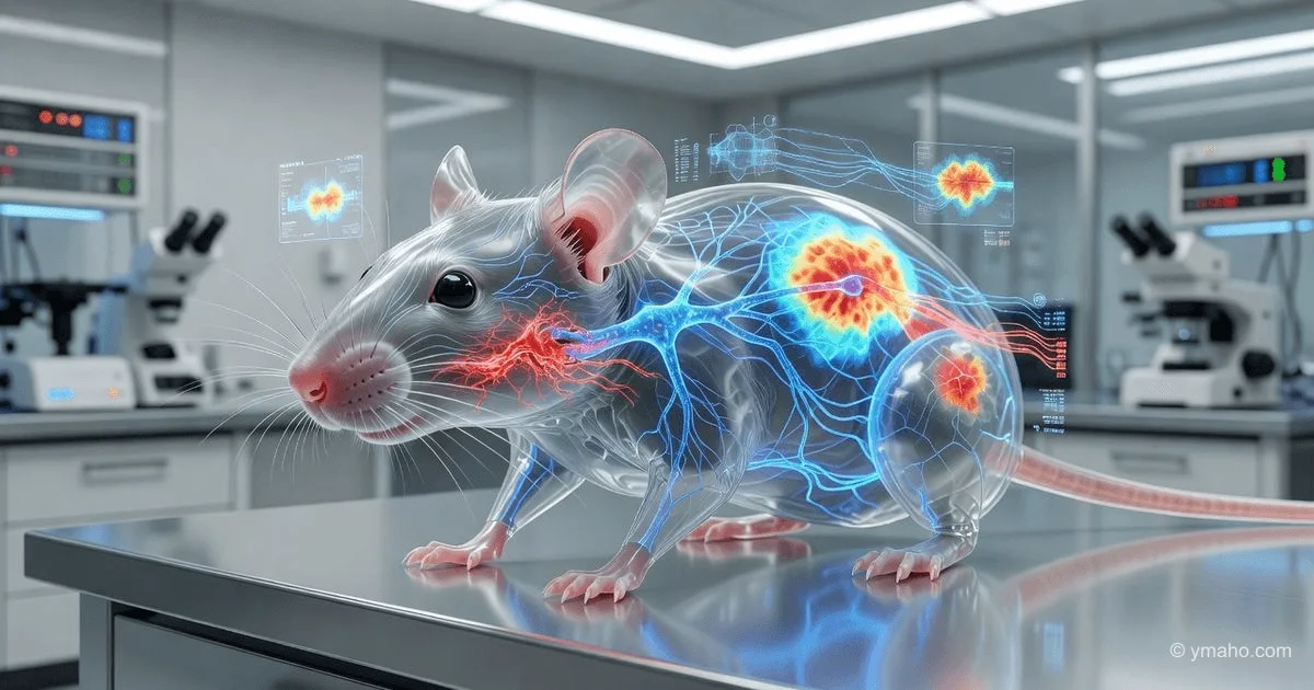
Here's how the technology works. First, mice are made optically transparent using the vDISCO method — this allows light to pass through the entire body, including bones and muscles, without scattering. Then, nerves and immune cells are labeled with fluorescent proteins (Uchl1-eGFP for nerves, Cd68-eGFP for macrophages). Next, a special light-sheet fluorescence microscope (LSFM) scans the entire body at a resolution sufficient to distinguish individual axons. The data volume from one mouse is up to 50 terabytes.
And this is where MouseMapper comes in. It is not a single algorithm but an ensemble of three modules based on the VesselFM foundation model:
- Nerve-Module — segments nerve fibers throughout the body, distinguishing even the finest axonal branches. The model was fine-tuned on manually annotated VR data and achieved a Dice score of 0.75, which is an outstanding result for 3D nerve segmentation.
- Immune-Module — identifies individual immune cells and their clusters. It recognizes even cells in previously unseen tissues — liver, intestine, muscles — without retraining.
- Tissue-Module — automatically segments 31 types of organs and tissues, creating an anatomical "skeleton" to which data from the first two modules are anchored.
The key technical achievement is the use of a foundation model. VesselFM was originally trained on blood vessel segmentation, but since vessels and nerves have similar topology (long branching structures), the model was able to transfer knowledge to the new task. This allows MouseMapper to work on data from different microscopes, with different magnifications, and with different labeling methods (reporter mouse lines vs. antibodies) without retraining.
Timeline and Context
The technology underlying MouseMapper has evolved over the last 5-7 years, and understanding this timeline is critical.
2017-2019: The group of Ali Ertürk (then at the Max Planck Institute for Biomedical Research) develops the vDISCO method — a tissue clearing technology that makes a mouse completely transparent for light microscopy. This was the first step: now the entire mouse could be "seen," but interpreting 50 TB of raw data was impossible.
2020-2022: The first attempts at automatic segmentation of organs and nerves emerge, but they are primitive — each organ requires a separate model, and nerves are often confused with vessels. Papers are published in specialized microscopy journals but do not reach the scale of Nature.
2023: VesselFM is released — a foundation model for blood vessel segmentation developed by the Ertürk group. This is a breakthrough: a model trained on tens of terabytes of data can segment vessels in any tissue, from any microscope, without retraining. The authors realize that the same architecture can be adapted for nerves.
2024: The team begins work on MouseMapper. According to the paper, they collected and annotated in VR about 84 subvolumes of size 300x300x300 voxels and 8 subvolumes of size 1000x1000x1000 voxels — a colossal effort to create ground truth that took at least a year.
Key date — May 19, 2026: Online publication in Nature. Interestingly, the paper appears in the main Nature journal (impact factor > 60), not in Nature Methods or Nature Biotechnology. This is significant because Nature rarely publishes purely technological works — they require biological insight. And they got it: the discovery of trigeminal nerve damage was sufficient for publication in the flagship journal.
Who Wins and Who Loses
Winner #1: Helmholtz Munich and LMU Munich as technology hubs.
Creating MouseMapper is a bid for global leadership in systems biology. Helmholtz Munich has traditionally been strong in metabolic diseases (diabetes, obesity). Now they have an exclusive tool that they can use to study other diseases — cancer, neurodegeneration, autoimmune disorders. This will attract tens of millions of euros in grants over the next 2-3 years.
Winner #2: Researchers in peripheral neuropathy.
Before MouseMapper, it was believed that obesity causes neuropathy only in the legs (diabetic polyneuropathy). The discovery of damage to the trigeminal nerve — the first cranial nerve — completely changes this view. It turns out that obesity can affect facial sensitivity, chewing, and even reflexes. For neurologists, this opens a new field of research.
Winner #3: Companies involved in high-throughput microscopy (Zeiss, Leica).
MouseMapper uses light microscopes with custom optics. Standard commercial systems cannot handle such data volume and resolution. Zeiss and Leica are likely already in talks with Helmholtz Munich to create a "packaged" solution — microscope + AI analytics — for pharmaceutical companies. The market for such systems could reach $200-300 million per year.
Loser #1: Traditional histology laboratories.
Organ biopsy followed by histology and manual cell counting is a multi-billion dollar market. MouseMapper offers an alternative: "biopsy" of the entire organism without cutting. It cannot completely replace histology (organelle-level detail still requires microscopy), but for screening systemic drug effects, the AI approach will be more efficient and cheaper.
Loser #2: Researchers without AI and computational biology skills.
MouseMapper requires a computing cluster (model training on 50 TB of data on 8-16 GPUs A100/H100) and expertise in foundation models. Small labs without such resources will not be able to reproduce or adapt the method. This deepens the gap between "hubs" and "periphery" in biomedical science.
What the Media Isn't Saying
Non-obvious insight #1: MouseMapper found trigeminal nerve damage by accident — it was an unintended discovery, not a hypothesis-driven experiment.
Here lies the most important point that press releases omit. The team did not set out to "find facial nerve damage in obesity." They simply ran MouseMapper on data from mice on a high-fat diet and looked at what the algorithm would show. And the AI "spit out" the trigeminal nerve as the area with maximal structural changes.
This changes the paradigm of scientific discovery. In traditional biology, we formulate a hypothesis, design an experiment, collect data. In the AI era, we can do the opposite: collect all data, run it through an algorithm, and see what we never thought to look at. This is "unbiased biology," and it has already yielded its first fruit.
Non-obvious insight #2: The effect on the nerve was colossal — 60% fewer endings.
Quantitatively, the changes are striking: in obese mice, the trigeminal nerve had 60% fewer branches, branch points, and nodes. Behavioral tests confirmed that the animals responded less to whisker (vibrissae) touch. So this is not about "mild functional decline" but about severe sensory deficit. Proteomic analysis of the trigeminal ganglion revealed changes in over 200 proteins, especially those involved in axonal remodeling and complement pathways.
Most shocking: when analyzing trigeminal ganglion samples from obese humans, the same molecular changes were found. This means the mouse model is not lying — in obese people, the facial nerve is actually damaged. How many obese patients have reduced facial sensitivity and don't even suspect it's related to their weight?
Non-obvious insight #3: The data is fully open — this is a strategic move, not altruism.
The authors announced that all 3D mouse atlases are available online. This is not just "sharing results." It is a way to become the standard. If thousands of labs start using MouseMapper and upload their atlases in the same format, Helmholtz Munich will become the center of the universe for systems biology. Competitors will be forced to join their ecosystem or create their own — which would take years.
What is missed about cost: Processing one mouse involves 50 TB of data. GPU cluster computations require at least 48-72 hours. Even at cloud rates ($2-4 per hour for an A100), one mouse costs $500-1000 just for computation, plus the cost of microscopy, tissue clearing, and reagents. MouseMapper is a technology for wealthy labs, not for routine screening.
Forecast: Next 30 Days and 90 Days
Next 30 days (until July 7, 2026):
Expect at least three pharmaceutical companies — Novo Nordisk (leader in obesity treatment), Eli Lilly (with their tirzepatide), and Pfizer (developing their own analogs) — to contact the Ertürk group for validation of MouseMapper on their animal models. If their drugs not only reduce weight but also restore the trigeminal nerve, this will become a powerful marketing argument: "Our medicine is not just for obesity, but also for facial sensitivity."
Concurrently, preprints will appear on bioRxiv attempting to adapt MouseMapper for other diseases — most likely for Alzheimer's disease models (where peripheral nerve damage also occurs) and for metastatic cancer (where macrophages infiltrate various organs). Given that the immune module already shows clusters of Cd68+ cells in the liver and adipose tissue, this could be used to study the metastatic microenvironment.
Next 90 days (until September 2026):
The most important shift will occur in commercialization. Ertürk and Schefer are likely already discussing creating a spin-off with the working name "OmniMapper" or "SysBioAI." Given the Nature publication, a seed round could reach $15-25 million from venture capital funds specializing in AI in biotech — for example, Andreessen Horowitz Bio or Khosla Ventures.
Also within 90 days, negotiations will begin with the Chan Zuckerberg Initiative (CZI). CZI invests billions in creating human cell atlases. MouseMapper is an ideal tool for scaling these atlases to the whole-organism level. A grant of $5-10 million is likely to be announced for adapting MouseMapper for human tissues.
Finally, I expect the Helmholtz Munich group to file a patent not on the algorithm itself (it is already open source), but on a "method for diagnosing trigeminal neuropathy associated with obesity using 3D imaging." This is a method-of-use patent that could be licensed to medical device manufacturers (e.g., Siemens Healthineers) to create non-invasive diagnostic systems.
Brief summary: MouseMapper is not a tool for studying obesity. It is proof that AI can "generalize" the anatomy of a whole organism just as GPT generalizes language. The next goal is to create a "digital twin" of a mouse, where any disease can be modeled before conducting a real experiment. And when that day comes, 70% of preclinical studies will become simulations, and mouse colonies will shrink by an order of magnitude. The question is not whether this will happen. The question is how many years it will take to scale MouseMapper from 50 TB per mouse to 500 TB per human.
— Editorial Team