Revolution in Neuroscience: A Protein Created to Listen to the Brain's "Hidden Language"
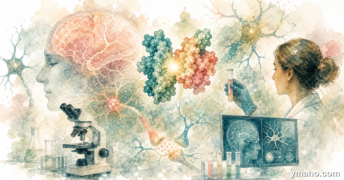
Researchers from the Allen Institute and the Janelia Research Campus published a paper in Nature Methods on the creation of a highly sensitive protein, iGluSnFR4 (a glutamate sensor), that allows visualization of incoming signals between neurons. This breakthrough opens the way to understanding the mechanisms of Alzheimer's disease, schizophrenia, and epilepsy at the synaptic level.
Analytical Review: iGluSnFR4 — A Breakthrough in Neuroscience or Just Another Expensive Tool?
Analysis Date: May 29, 2026
[The Gist]: What's Really Happening
On the surface, it's a nice story from the "science moves forward" section. Scientists from the Allen Institute and the Janelia Research Campus created the iGluSnFR4 protein, which allows real-time visualization of incoming signals between neurons. A publication in Nature Methods. Finally, we can "hear" the brain's hidden language. It opens the way to understanding Alzheimer's, schizophrenia, and epilepsy.
It sounds like a revolution. And it is indeed an important instrumental breakthrough. But only for a narrow circle of specialists.
The main non-obvious insight that isn't in the headlines:
iGluSnFR4 is not a breakthrough in treatment. It's a breakthrough in fundamental research. The difference between "we saw a signal in a mouse neuron" and "we cured Alzheimer's in a human" is a chasm 10-15 years long and billions of dollars deep. All claims about "new paths for treatment" are standard grant application phrasing, not clinical reality. iGluSnFR4 will become a standard tool in a few hundred neurobiology labs worldwide. But patients won't see its benefits until at least 2040.
Google AdInline article slot
Timeline and Context
2015 — First generation iGluSnFR from the group of Loren Looger (HHMI Janelia). A sensor based on a bacterial periplasmic glutamate-binding protein and green fluorescent protein. Revolution: for the first time, glutamate could be seen in living neurons. But the problem: slow — 2-3 ms, insufficient for tracking fast synaptic events at 100 Hz.
2019 — Emergence of ultra-fast variants iGlu_u and iGlu_f with a dissociation constant of 600 µM. Dissociation became 6 times faster, allowing visualization of glutamate clearance from the synaptic cleft during 100 Hz stimulation. But sensitivity left much to be desired — the signal from a single vesicular release was too weak.
December 2025 — Publication in Nature Methods of the fourth generation. The authors screened 3,365 variants of iGluSnFR3 in neuronal culture and selected the two best for in vivo testing in the mouse visual cortex. Results:
- iGluSnFR4f (fast deactivation): activation time <2 ms, deactivation time 25.9 ms. Capable of tracking fast synaptic events, including whisker stimulation in mice at 20 Hz.
- iGluSnFR4s (slow deactivation): deactivation 152.7 ms. Ideal for recording large populations of synapses at low frame rates (30 Hz). At this frame rate, it detects 9 times more synapses than iGluSnFR3.
Real numbers that impress insiders:
- Signal amplitude per single action potential — significantly higher than iGluSnFR3 (difference not disclosed, but clearly statistically significant).
- Sensitivity — single vesicular release detected in vivo.
- Spatial specificity — signal localized at the level of individual dendritic spines, minimal crosstalk between neighboring synapses.
- Photostability — 1 hour of continuous imaging at 100 Hz, retaining 75-87% of signal.
Why this matters for fundamental science: Before iGluSnFR4, neurobiologists could only measure outgoing signals (action potentials) or structural connections. Incoming signals — what thousands of synapses "say" to the neuron — were a black box. iGluSnFR4 allows, for the first time, visualization of synaptic activity patterns that drive a neuron's decision to "fire or not fire." It's like moving from hearing individual words to understanding the grammar of a language.
Who Wins and Who Loses
Absolute winner — academic neuroscience. Labs studying synaptic plasticity, learning and memory, sensory processing, and neurodegenerative diseases now have a tool to see what was previously invisible. In particular, the group of David Kleinfeld from UC San Diego (one of the co-authors) is already using iGluSnFR4f to study sensory signal processing in the barrel cortex during whisker stimulation.
Winner — HHMI Janelia Research Campus and Allen Institute. This publication strengthens their position as world leaders in developing neuroscience tools. iGluSnFR4 is already available through Addgene (a non-profit plasmid repository). The next 5-10 years of thousands of citations in Nature/Science/Neuron are guaranteed.
Winners — drug development companies for neurodegenerative diseases (Biogen, Eisai, Roche, Eli Lilly). They now have a tool for target validation in animal models. Instead of extrapolating drug effects from indirect signs (behavior, neuronal survival), they can directly see how their molecule affects glutamatergic synaptic transmission. This will accelerate the preclinical phase — but not the clinical one.
Loser — old methods. Electrophysiology (patch-clamp, multi-electrode arrays) remains the gold standard for measuring neuronal activity with high temporal resolution. But it provides no spatial information — you know the neuron fires, but you don't see which synapses activated it. iGluSnFR4 won't replace electrophysiology, but will complement it. However, grant applications relying solely on old methods will look less competitive.
Non-obvious loser — two-photon microscopy as a bottleneck. iGluSnFR4 requires two-photon imaging at frame rates of 30-500 Hz. This equipment costs $500,000-$1,500,000 per setup and requires highly skilled personnel. iGluSnFR4 does not democratize neuroscience — it will widen the gap between the "top-10" labs with such equipment and everyone else.
What the Media Isn't Saying
First and most important. All news shouts "breakthrough in treating Alzheimer's, schizophrenia, epilepsy." But look at the experimental design. iGluSnFR4 was tested on healthy mice shown visual stimuli or whisker stimulation. Not a single experiment on disease models. Not a single drug was tested. Claims about "new paths for treatment" are wishful thinking wrapped in scientific rhetoric.
Second. The technology works in mice, not humans. For human application, you need: (1) a safe way to deliver the iGluSnFR4 gene into human neurons (AAV — possible, but immune response), (2) two-photon microscopy through an intact human skull (technically very difficult, requires implantation of an optical window), (3) ethics committee approval for genetic modification of living human neurons. This will not happen in the foreseeable future. iGluSnFR4 will remain a tool for animal models.
Third. Even in mice, there are limitations. iGluSnFR4 requires membrane localization (NGR or PDGFR signal sequence). This means the sensor is embedded in the neuron's membrane. Expression of the sensor itself may affect synaptic function. The authors checked dendritic spine survival and found no differences from controls. But subtler effects (altered receptor deactivation time, changes in synaptic plasticity) are not ruled out.
Fourth. "Ultra-sensitivity" is marketing. The Nature Methods paper states that iGluSnFR4 detects single vesicular release. But in a real experiment, at physiological stimulation frequencies, signals from neighboring synapses overlap. Resolving single events is possible only at very low frequencies (1-5 Hz) or with pharmacological receptor blockade. Under natural conditions at 20-100 Hz, the signal becomes summated.
Fifth. iGluSnFR4 does not distinguish glutamate from different synapse types. Glutamate is glutamate everywhere. The sensor doesn't know whether the signal came from an excitatory synapse (AMPA/NMDA receptors) or astroglial transport. All it sees is extracellular glutamate concentration. Data interpretation requires caution.
Forecast: Next 30 Days and 90 Days
30 days:
A surge of plasmid downloads from Addgene will begin. I expect that within a month, 200-300 labs worldwide will order iGluSnFR4 constructs. Concurrently, the first preprints using iGluSnFR4 in new contexts will appear — for example, in Alzheimer's disease models (APP/PS1 mice) or traumatic brain injury.
Nature Methods will likely organize a virtual symposium or webinar with the authors. The key question from the community: what is the signal variability between different animals and different AAV batches? The authors claim they "see no noticeable differences," but independent replication will tell.
90 days:
First replication studies from independent groups. If iGluSnFR4 is easily reproducible, it will become the standard. If problems arise (low expression in certain neuron types, phototoxicity during prolonged imaging), development of iGluSnFR5 will begin.
Second event: release of protocols for specific applications. The authors will publish detailed instructions for using iGluSnFR4 in different areas: cortex (layers 1-4), hippocampus (CA1), midbrain (photometry via optical fiber). This will lower the entry barrier for labs without experience with genetically encoded sensors.
Long-term forecast (2026-2030):
iGluSnFR4 will become a standard tool in neuroscience, like GFP or GCaMP (calcium indicator). By 2028, it will be used in 50-70% of publications on synaptic plasticity and sensory processing. By 2030, variants for other neurotransmitters (dopamine, serotonin, GABA) based on a similar platform will appear.
But the path to the clinic — if possible at all — will take decades. Even for diagnostics (not therapy), invasive delivery and imaging are required. The only scenario where iGluSnFR4 could be used in humans is ex vivo analysis of brain tissue biopsies (after epilepsy or tumor surgery). And even then, with enormous ethical and regulatory barriers.
**And finally: don't believe headlines about a "breakthrough in treatment." iGluSnFR4 is a microscope, not a drug. It will let us see how neurons communicate. But seeing is not fixing. We still can't cure Alzheimer's despite decades of studying amyloid plaques and tau protein. iGluSnFR4 will add a new chapter to understanding pathogenesis. But the distance to therapy is enormous.
— Editorial Team